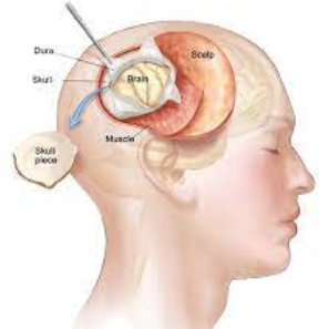
Neuro-endoscopy is a minimally invasive surgical technique that
involves the use of an endoscope to visualize and treat conditions within the
brain, ventricles, or subarachnoid space. The endoscope is a thin, flexible
tube with a light source and a camera at its tip, allowing neurosurgeons to
access and navigate through narrow or deep structures in the brain. Neuro-endoscopy
is used for diagnostic purposes as well as for treating various neurological
conditions.
Purpose:
Visualization and Treatment: Neuro-endoscopy enables surgeons to visualize and
access areas within the brain and surrounding structures.
Minimally Invasive: The procedure is considered minimally invasive
compared to traditional open surgery, as it typically involves smaller
incisions.
Indications for Neuro-endoscopy:
Ventricular Conditions: Neuro-endoscopy is commonly used for procedures
involving the ventricular system, such as the third ventricle or lateral
ventricles.
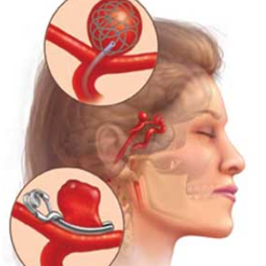
Cyst Removal: Neuro-endoscopy can be used to remove cysts or
tumors located within the brain.
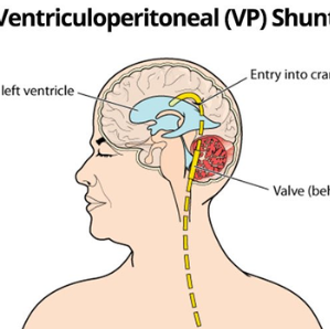
Hydrocephalus Treatment: In some cases of hydrocephalus (accumulation of
cerebrospinal fluid), neuro-endoscopy may be employed to create a communication
pathway or perform fenestration to relieve fluid buildup.
Tumor Biopsy: Neuro-endoscopy can be used to obtain biopsies of
tumors within the brain.

Neuro-endoscopy


.png)