Coronary Angiography is a medical procedure used to visualize the coronary arteries, which
supply blood to the heart muscle, and to assess the presence of blockages,
narrowing, or other abnormalities within these arteries. It is a crucial
diagnostic tool for evaluating and diagnosing coronary artery disease (CAD),
which can lead to reduced blood flow to the heart and may result in chest pain
(angina) or more severe conditions, such as heart attacks.
Here's how
coronary angiography is typically performed:
Preparation: Before the
procedure, the patient is prepared by having an intravenous (IV) line inserted
to provide medications and fluids. Electrocardiogram (ECG) leads are attached
to monitor the heart's electrical activity. Sedation or anesthesia may be
administered to help the patient relax and remain comfortable during the
procedure.
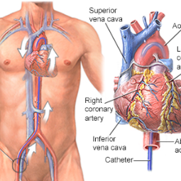
Access: Coronary
angiography is typically performed through the femoral artery in the groin or
the radial artery in the wrist. The healthcare provider cleans the area and
numbs it with a local anesthetic. A thin, flexible tube called a catheter is
then inserted into the artery.
Guidance: The
catheter is carefully advanced through the vascular system to the heart and coronary
arteries. It is guided using X-ray imaging to ensure proper placement.
Contrast Dye Injection: Once the catheter is in position, a contrast dye is injected through the
catheter directly into the coronary arteries. The dye is visible on X-ray,
allowing the healthcare provider to see the blood vessels and any blockages or
abnormalities.
Imaging: Continuous
X-ray imaging is used to capture a series of images of the coronary arteries as
the contrast dye flows through them. These images provide a detailed view of
the arteries, highlighting any areas of narrowing, blockages, or other issues.
Assessment: The
cardiologist reviews the images and assesses the condition of the coronary arteries.
They can identify the severity and location of any blockages or stenosis
(narrowing).
Decision-Making: Based on the findings, the cardiologist may decide on further treatment
options, such as angioplasty and stent placement (percutaneous coronary
intervention, or PCI), or they may recommend other therapies or lifestyle
changes.
Closure: After the
procedure, the catheter is removed, and the access site (groin or wrist) is typically
sealed with a special device or manual pressure to prevent bleeding. The
patient is monitored for a few hours to ensure stability.
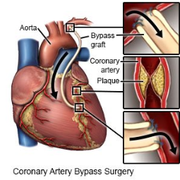
Coronary angiography is considered the gold standard for diagnosing
coronary artery disease because it provides precise information about the
extent and location of blockages or narrowing in the coronary arteries.
Depending on the findings, the cardiologist can make informed decisions about
the most appropriate treatment, which may include angioplasty and stent
placement or, in some cases, coronary artery bypass surgery (CABG).
While coronary angiography is generally safe, it carries some risks,
such as bleeding or damage to the blood vessels. The benefits of accurate
diagnosis and treatment planning typically outweigh these risks for patients
with suspected or known CAD. The healthcare team will discuss the procedure's
risks and benefits with the patient before proceeding.


.png)